


How cyanobacterial cells communicate reveals the importance of cellular architecture
How cells are arranged within cyanobacterial filaments affects how they communicate with one another. New research from the Department of Ecology, Environment and Plant Sciences (DEEP) at Stockholm University provides new insights into how cellular architecture shapes communication in multicellular bacteria.
The study, published in Applied and Environmental Microbiology, investigated how cells communicate in the filamentous facultative symbiotic cyanobacterium Richelia rhizosoleniae. The researchers found that the way cells are organised within a filament influences how substances move between them, providing new insights into how multicellular organisation works in bacteria.
Communication through tiny connections
Unlike most bacteria, filamentous cyanobacteria can function as coordinated communities of many connected cells. Molecules can pass directly between neighbouring cells through specialised structures called septal junctions.
These connections are particularly important in cyanobacteria that contain different types of cells with specialised roles. For example, some cells called heterocysts are specialised for nitrogen fixation, converting atmospheric nitrogen into a form that can be used by other cells. Critical to heterocysts are the provision of energy in the form of sugar from the photosynthesis performed by their neighboring vegetative cells.
But how does the organisation of these different cells affect the exchange of molecules within a filament?
To investigate this, researchers Sepehr Bardi, Mercedes Nieves-Morión and. Rachel Foster compared Richelia rhizosoleniae strain SC01 with the model cyanobacterium Anabaena spp. 7120. They also included a mutant Anabaena strain in which communication is impaired.
The researchers used two live cell imaging techniques and a confocal microscope housed at IFSU to track how fluorescent molecules are exchanged between cells (heterocysts and vegetative) and also along filaments. They also used transmission electron microscopy to examine the tiny pores connecting the cells.
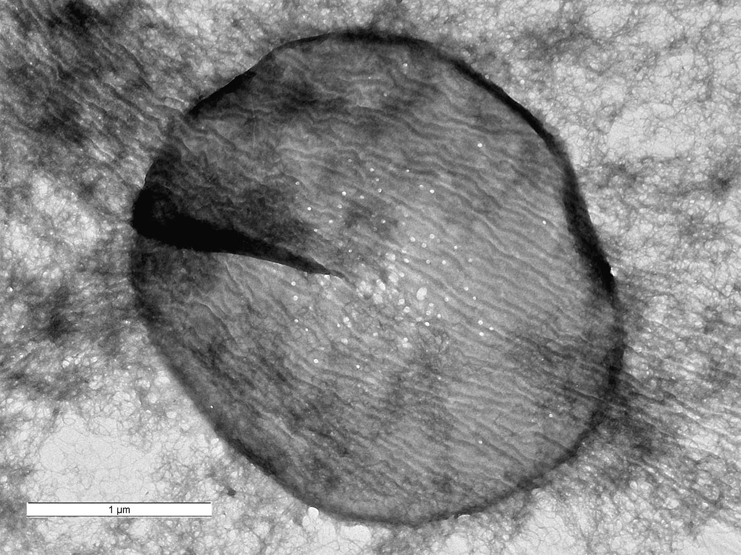
Electron micrograph of the septal disc of Richelia SC01 with visible nanopores. Image: Juan Luis Ribas of the Servicio de Microscopía, Universidad de Sevilla, Seville, Spain.
The shape of the filament matters
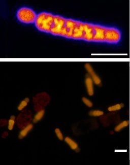
Richelia SC01 under the microscope. The top image shows a free-living filament, tapering towards a specialised cell called a heterocyst. The bottom shows Richelia living attached to Chaetoceros diatoms. Richelia fluoresces yellow and the diatom chloroplasts red. Scale bars are approximately 10 μm. Top image: Sepehr Bardi and IFSU; bottom image: Rachel Foster.
Richelia has a distinctive filament structure that becomes narrower towards one end, with a specialised heterocyst at the end of the filament. The researchers found that this pronounced tapering did not simply result in a one-way flow of molecules. Instead, vegetative cells in Richelia showed relatively symmetrical exchange with their neighbours.
The experiments showed that Richela and Anabaena had similar overall rates of molecular exchange, despite having important differences in their cellular organisation.
This contrasts with Anabaena, where the exchange between cells was more asymmetric despite a more uniform filament structure and possessing heterocysts in the middle of their filaments. Lead author of the study, Sepehr Bardi, explains:
The results suggest that the organisation of a filament alone cannot explain how substances move between its cells. Cell identity, cell shape and the organisation of the connections between cells all contribute to the communication pattern.
The researchers also found differences in the number of tiny pores, or nanopores, in the septa between Richelia cells. The pores were similar in size to those found in Anabaena, but their numbers varied considerably. This may help explain some of the differences in how molecules are exchanged between cells.
Understanding bacterial multicellularity
For organisms that live as filaments, communication between cells is essential. Cells need to exchange metabolites and other molecules while carrying out different functions, allowing the filament to behave as a coordinated whole.
The new findings show that this coordination is more complex than simply having channels that connect neighbouring cells. Instead, communication emerges from the interaction between the physical structure of the filament and the specialised properties of individual cells.
The study also demonstrates how combining different live cell imaging can provide a quantitative picture of communication within and along bacterial filaments. This can help researchers better understand how multicellular organisation has evolved in microorganisms and how different cell types coordinate their activities. Answering the questions on what's ahead, co-author Rachel Foster explains:
I think another exciting next step is to study the communication dynamics in a symbiotic life style. This strain has been in isolation for many years, but recently we found a host diatom that it actively attaches to under certain conditions… the tricky part will be setting up the live cell imaging while its symbiotically attached!
Read more
Access the publication in Applied and Environmental Microbiology via the below link:
Last updated: 2026-09-24
Source: Department of Environmental Science,Department of Ecology, Environment and Plant Sciences