


Immune molecules play a limited role in ALS progression
Immune-related molecules change in motor neurons affected by amyotrophic lateral sclerosis (ALS), but they do not appear to be major drivers of disease progression. The findings, published by researchers at Stockholm University, provide new mechanistic insights into disease pathology in ALS.

Eva Hedlund and Melanie Leboeuf. Photo: Anette Gärdeklint Sylla
“One of the central questions in ALS is why some motor neurons die while others survive. Our study examined whether immune-related molecules could explain this selective vulnerability, but we found that they are only one small piece of a much larger puzzle,” says Professor Eva Hedlund, who led the study together with Professor Staffan Cullheim at Karolinska Institutet.
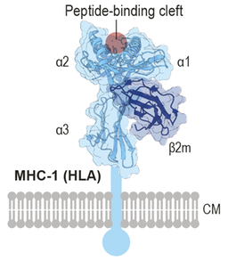
Schematic of the MHC-I molecule. Image: E. Hedlund and M. Karlen
The researchers focused on the major histocompatibility complex I (MHC-I), a group of molecules best known for their role in the immune system but also important for normal nervous system function. Previous studies have suggested that reduced levels of MHC-I components may make motor neurons more vulnerable in ALS.
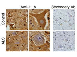
Antibody staining against HLA protein in post mortem donor tissues shows a significant decrease in ALS end-stage patient spinal motor neurons. Image: Melanie Leboeuf
Motor neurons try to counteract disease
The team found that the largest and most vulnerable spinal motor neurons naturally express lower levels of HLA, a key component of MHC-I. In tissues from ALS patients, HLA protein levels were reduced even further, while they remained preserved in disease-resistant oculomotor neurons that control eye movements.
While HLA protein levels declined, ALS spinal motor neurons increased their mRNA expression of certain HLA transcripts and of β2-microglobulin (β2m), another pivotal component of MHC-I. This indicates an attempted compensatory response that fails to restore protein levels.
“The findings suggest that vulnerable motor neurons actively try to counteract disease-associated stress but that this response is ultimately insufficient,” says Dr Melanie Leboeuf, the study's first author.
*Human leukocyte antigens (HLAs) are cell-surface proteins that present peptides to T cells, enabling the immune system to distinguish healthy cells from infected or abnormal cells.
*Beta-2 microglobulin (β2m) are small proteins found on the surface of most cells in the body, helping the HLA proteins to work properly. Together HLAs and b2m constitute the so-called MHC-I complex.
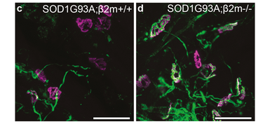
Removing the b2m gene in ALS mice resulted in partial preservation of synapses between motor neurons and muscle in particular muscles. Image: Melanie Leboeuf
A complex disease process
To investigate whether β2m influences disease progression, the researchers removed the gene in a mouse model of ALS. The researchers found that deleting β2m helped preserve nerve connections to specific muscles, but the effect was limited and did not improve overall motor function or lifespan of the mice. Unexpectedly, the team discovered that mice lacking the β2m gene exhibited marked astrocyte activation, even in the absence of motor neuron disease.
*Astrocytes are cells in the nervous system that play an activate role in ALS and appear to attack motor neurons in the disease.
Loss of β2m also resulted in further increased astrocyte activation in ALS mice, yet without worsening disease progression. This suggests that β2m normally contributes to regulating astrocyte reactivity. It also demonstrates that activation of astrocytes alone is insufficient to drive motor neuron degeneration, even when neurons lack functional MHC-I on their surface. It also suggests that once a threshold in astrocyte activation is reached, further reactivity does not exacerbate disease.
“We found that beta2-microglobulin clearly influences how motor neurons and their environment respond to disease yet removing it had remarkably little effect on overall disease progression. That tells us it is not a major disease modifier in ALS,” says Melanie Leboeuf.
The findings show that while MHC-I-related molecules are dynamically regulated in ALS, they are unlikely to be key drivers of disease progression. Instead, they appear to represent one small part of a much more complex disease process, providing researchers with a clearer picture of the biological mechanisms underlying ALS.
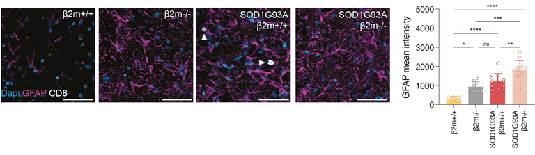
Removing b2m resulted in an activation of astrocytes in healthy mice and even further in ALS mice. Image: Melanie Leboeuf
More about the study
To read the full study, please see Springer Nature's website:
Last updated: 2026-08-14
Source: Department of Biochemistry and Biophysics